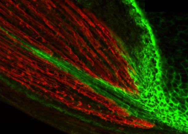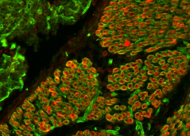
LAMA2-congenital muscular dystrophy (LAMA2-CMD, also called MDC1A) is caused by mutations in the LAMA2 gene, which encodes the α2 chain of laminin 211 (LN211), the most important laminin isoform in mature skeletal muscle. A basement membrane matrix containing LN211 normally ensheaths all skeletal muscle fibers. It is generally believed that the absence of LN211 around myofibers in LAMA2-CMD patients causes a constant stress on these cells, which progressively damages them, inducing muscle wasting, inflammation and fibrosis. This environment in turn is thought to hamper regeneration, leading to a vicious circle of disease progression.
Within an AFM funded research project (nº 19959), we used the dyW mouse model for LAMA2-CMD to trace the onset of the disease and found that it starts during fetal muscle development, without any signs of muscle inflammation and fibrosis (Nunes et al., Hum Mol Gen 26:2018-33, 2017). More specifically, fetal dyW muscles display a precocious reduction in the number of muscle stem cells (MuSCs) and differentiated myoblasts, with a consequent failure in muscle growth. The timing of the defect (detected between E17.5 and E18.5) correlates with a stage at which MuSCs have just entered their niche under the basement membrane of myofibers, which in the case of dyW mice (and LAMA2-CMD patients) does not contain LN211.
Our studies within the past year have addressed whether fetal MuSCs contribute to the construction of their niche and how they interact with their niche. Our unpublished data, which include an RNAseq analysis, in situ hybridization, in vivo analysis and isolation and culture of fetal MuSCs and myoblasts demonstrate that these cells normally produce laminins, including LN211. Moreover, they express both the α7β1 integrin and dystroglycan when they enter their niche pointing to an active interaction between the MuSCs/myoblasts and their niche at a time we see an impaired expansion of these cells in dyW fetuses.
Altogether our data raise the new possibility that the primary defect of dyW mice (and LAMA2-CMD patients) is a MuSC defect. A promising avenue for LAMA2-CMD therapy would be to address this primary defect, thus targeting the origin of the disease. However, the detailed mechanisms underlying the precocious reduction in the number of MuSCs and myoblasts in fetal dyW muscles must first be dissected out.
Here we propose to zoom in on the population of MuSCs and myoblasts in dyW fetuses. The aim is to dissect out their response to LN211-deficiency in three complementary and interlinked objectives, focusing on the critical period between E17.5 and E18.5. We have successfully isolated pure populations of MuSCs and myoblasts from fetal muscles and have characterized these cells in vitro. We have also processed isolated MuSCs/myoblast and processed them for RNAseq analysis where we compared the transcriptional profile of WT and dyW MuSCs/myoblasts at E17.5 and E18.5. We find that while WT MuSCs/myoblasts significantly change their transcriptional profile as they settle in their niche (i.e. from E17.5 to E18.5), dyW MuSCs/myoblasts, which do not have LN211 in their niche, fail to change as much. These data provide compelling indications that normally WT MuSCs/myoblast do indeed react to their niche and raise the possibility, which we intend to test further in Objective 1 in this proposal, that dyW cells behave differently.
Since we know MuSCs/myoblasts normally produce LN211, in Objective 2 we ask if it is the LN211 MuSCs/myoblasts produce or the LN211 that myofibers produce that is most important for their niche construction. One way to address this outstanding question is to decellularize fetal WT and dyW muscle and analyze how isolated MuSCs and myoblasts seeded into WT and dyW matrices behave in these matrices within and across genotypes. These experiments will inform us about the relative importance of MuSC/myoblast-derived LN211 versus the LN211 present around myofibers in the WT decellularized ECM but absent in the dyW decellularized ECM. We have already set up the decellularization protocol for fetal WT and dyW muscles, have characterized these matrices and found that myogenic cells (C2C12 cells) invade and form myotubes in these matrices.
Finally, in Objective 3 this 3D culture assay will be used in parallell with 2D culture of isolated fetal MuSCs/myoblasts to validate the results from Objective 1. We will also design experiments to test the most promising genes/pathways that come out of Objective 1 in vitro.
This project represents a completely new and innovative approach to the study of LAMA2-CMD pathogenesis. It will be pursued by a multidisciplinary team with a proven scientific and technical track record in the issues addressed. Through this collaborative effort, we expect to contribute to the characterization of the primary defect of LAMA2-CMD and define possible targets for therapeutic development.

